
Dental photogrammetry has attracted increasing attention in implant dentistry because it can capture the three dimensional positions of implants with high accuracy. A photogrammetry 3D scanner uses photographic information and reference markers to calculate spatial relationships, providing an alternative to conventional impressions and some intraoral scanning workflows.
However, like any digital technology, photogrammetry is not suitable for every clinical situation. Understanding its limitations is important when evaluating whether a photogrammetry dental scanner fits a particular implant workflow. Recent research indicates that photogrammetry can provide reliable implant position data, particularly for full arch implant cases, while also highlighting limitations related to data scope, workflow integration, equipment requirements, and clinical conditions.
Photogrammetry Does Not Capture Every Type of Oral Data
One of the most important limitations of conventional dental photogrammetry is that it primarily records the three dimensional position of implants or scan markers. It does not necessarily capture the complete oral environment, including teeth, gingiva, occlusal relationships, and soft tissue.
This means photogrammetry alone may not provide all the digital information required for designing a restoration. A separate intraoral scan or another impression method may still be necessary to collect soft tissue and occlusal data. These datasets then need to be aligned during the digital workflow, creating another stage where discrepancies can potentially occur.
For clinicians and dental laboratories, this is an important consideration. A technology that accurately records implant positions is useful, but its practical value also depends on how easily the data can be combined with other digital records.
Accuracy Can Depend on the Clinical Workflow
Accuracy is often one of the first concerns when selecting a photogrammetry 3D scanner. Research generally supports the ability of photogrammetry to provide accurate implant position information, but results can vary according to the system, scan body design, number and distribution of implants, scanning method, and data processing workflow.
Complete arch cases can be particularly demanding. When multiple implants are distributed across a large arch, the system must establish precise spatial relationships between reference markers. In clinical environments, posterior areas may also be more difficult to capture because of interference from surrounding oral structures. Some research has identified the possibility of cumulative errors as the system records relationships between successive scan bodies.
Therefore, rather than considering photogrammetry inherently accurate or inaccurate, users should evaluate the complete system and workflow used to acquire and process the data.
Equipment and Workflow Requirements Matter
Another limitation is the additional equipment and workflow requirements associated with traditional photogrammetry. Specialized cameras, reference markers, scan bodies, and compatible software may be required. Staff also need to understand how these components should be positioned and how the resulting data should be integrated into CAD/CAM workflows.
For practices already using digital dentistry, introducing another independent system can create workflow complexity. The question is therefore not simply whether a photogrammetry dental scanner can capture implant positions, but whether it can fit efficiently into the existing digital process.
This is one reason integrated solutions are becoming increasingly relevant. Instead of treating implant position capture, intraoral scanning, and soft tissue acquisition as completely separate steps, an integrated approach can reduce the need for repeated data acquisition and manual alignment.
Conventional Photogrammetry Has a Restricted Scanning Scope
Traditional photogrammetry is particularly useful for implant position recording, but its restricted scanning scope can limit its application beyond implant dentistry. A systematic review noted that photogrammetry has shown strong potential as an implant coordinate transfer system, while its use as a complete three dimensional scanner has historically required further development.
This distinction is important when comparing different technologies. An intraoral scanner is generally expected to capture broader surface information, while photogrammetry focuses on accurately establishing spatial coordinates.
For full arch implant cases, this focused capability can be advantageous. For workflows requiring extensive surface data, however, users may still need another scanning technology.
How Integrated Photogrammetry Can Reduce These Limitations
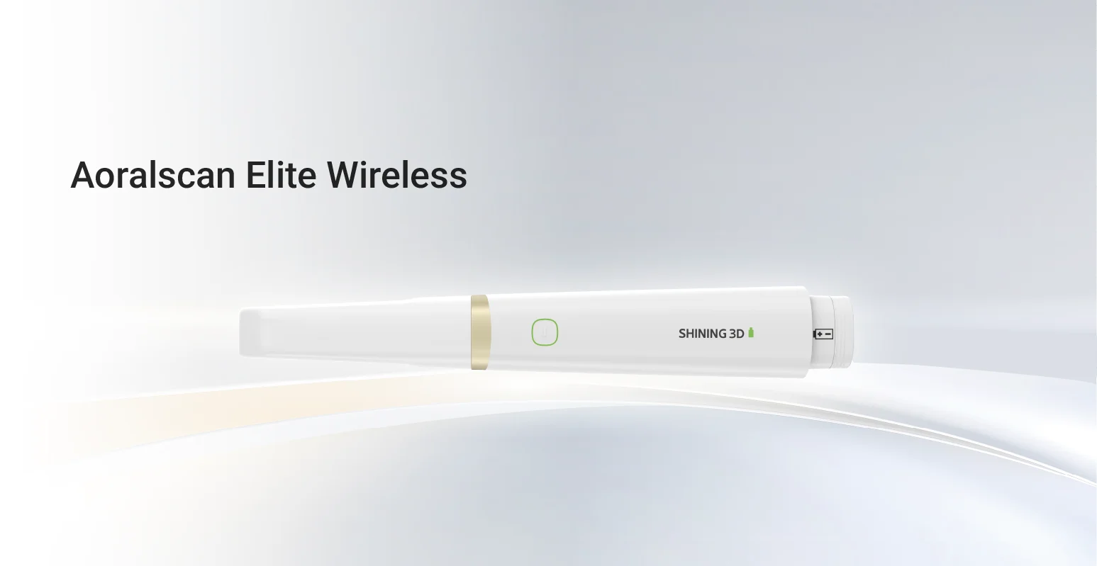
The development of intraoral photogrammetry addresses some of the practical limitations associated with traditional photogrammetry. SHINING 3D DENTAL integrates intraoral photogrammetry with intraoral scanning technology, allowing implant positions and angles to be acquired alongside detailed information about teeth and gingival soft tissue.
Its IPG approach is designed for applications such as edentulous implant cases and partial edentulism, using high precision encoded scan bodies to acquire implant information while also collecting intraoral data. This integration can reduce the need to manage completely separate scanning processes and may simplify the transition from clinical data acquisition to digital design.
For users evaluating a photogrammetry dental scanner, this type of integrated workflow is worth considering because the practical limitation of photogrammetry is often not the ability to measure implant positions, but how that information is combined with the rest of the patient’s digital records.
Choosing the Right Photogrammetry Workflow
Dental photogrammetry should be evaluated according to the clinical indication rather than treated as a universal replacement for intraoral scanning or conventional impressions. For full arch implant rehabilitation, accurate implant position data can be particularly valuable. For cases requiring comprehensive surface, soft tissue, and occlusal information, an integrated scanning solution may provide a more complete workflow.
Current evidence suggests that photogrammetry can offer high accuracy for complete arch implant impressions, although more clinical research is still needed and study results vary according to methodology and system.
The key is to consider accuracy, scanning scope, workflow integration, equipment requirements, and compatibility with downstream CAD/CAM processes together.
Making Photogrammetry More Practical for Digital Implant Dentistry
Dental photogrammetry has clear advantages for recording implant positions, but its traditional limitations should not be overlooked. Restricted data capture, additional scanning requirements, workflow complexity, and dependence on system specific components can affect how efficiently the technology is used.
Modern solutions such as SHINING 3D DENTAL IPG demonstrate how integrating photogrammetry with intraoral scanning can address some of these challenges. By combining implant position and angle acquisition with intraoral data collection, the workflow can better support the requirements of digital implant treatment.
For practices and laboratories considering a photogrammetry 3D scanner, the most important question is therefore not simply how accurate the technology is, but whether the complete workflow provides the data, compatibility, and efficiency required for predictable digital implant restoration.